A Microfluidic Approach to the Brain
by Noah Pflueger-Peters
The flow of blood and cerebrospinal fluid (CSF) is critical to the brain. The brain has a limited energy reserve, so blood needs to bring oxygen and nutrients to neurons immediately upon activation, a phenomenon known as neurovascular coupling or functional hyperemia. CSF washes out metabolic waste generated by neurons while cushioning the brain and spinal cord from injury.
These important transport processes occur mostly in capillaries with a diameter of 5-8 micrometers, the smallest blood vessel in the brain. To microfluidic researchers like Chemical Engineering Associate Professor Jiandi Wan, this is an opportunity to play a significant role in discovering how the brain works.

Wan’s group studies transport phenomena in chemical and biological systems using microfluidic approaches. The team cultures stem cells and investigate the fluid dynamics of biological fluids on tiny scales in microfluidic devices, also known as labs-on-a-chip. Combined with live in vivo studies, these microchip-sized instruments give researchers a well-controlled experimental environment they can use to allow researchers to identify the definitive roles of these fluids.
“It’s amazing how fluid movement significantly impacts brain functions,” he said. “This is pure fluids and occurs on microscales, so I think it’s going to be a perfect area for microfluidics researchers to dig in to find out what’s going on.”
Fluids and the Brain
Wan is fascinated by the richness of fluid dynamics in biological systems. An example he mentions is how arterials, medium-sized blood vessels, have smooth muscle cells that allow them to constrict or dilate to control the direction of blood flow to different parts of the body.
“That means a really small change of diameter can dramatically change the flow rate,” he said. “It seems that our body knows that, and it’s how the heart pumps blood and how blood is distributed to our brain or the stomach.”
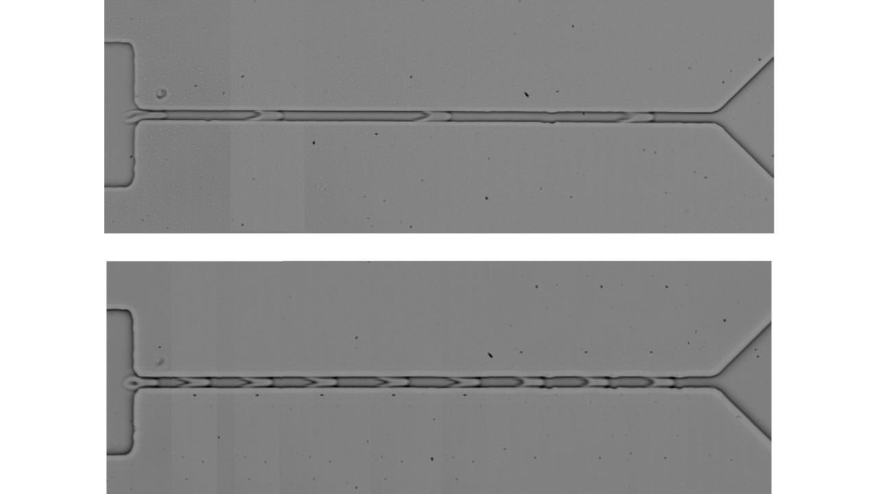
In two studies in 2016 and 2019, Wan’s team tested the claim that this phenomenon was responsible for functional hyperemia and found it was caused by red blood cells in the capillaries instead.
“We confirmed that functional hyperemia in the brain was initiated in the capillaries and the red cells play a huge role,” he said. “When neurons are activated, they consume oxygen, and red blood cells in our body can sense the oxygen change and become more deformable. That triggers the series of reactions upstream to induce dilation and increase blood flow.”
A Bright Future
Recently, Wan has become an evangelist for microfluidics in brain research. In a paper in the January 2022 issue of Chemical Reviews, Wan and his co-authors outline three brain research areas that microfluidics can impact—CSF, brain-on-a-chip devices and neuron-electronic devices.
Researchers recently discovered that CSF carries molecules excreted by neurons and that its flow can remove metabolic wastes and neurotoxins from the brain. However, the driving force behind CSF flow, the exact drainage pathways and how it’s produced in the brain remain debated. Wan thinks innovative engineering approaches like microfluidics can shed light on these fundamental questions.
One way to do this is brain-on-a-chip systems, which could serve as fully-functional micro-models of the brain outside of the body. Similar models have been built for other organs, but integrating cultured neurons, blood vessels and tissue cells in a microfluidic device that mimics living brain structure and function has not yet been achieved. If successful, Wan says researchers can study not only the fluids in the brain, but also the interactions between neurons, blood vessels and tissues cells in a controlled setting.
“I think it’d be really exciting to use microfluidics to develop an artificial brain in a microchip so you can track the neural activity and the fluids inside the mini-brain,” he added. “Ex vivo microfluidics studies also provide a unique advantage in terms of control because we can isolate key components, focus on that and then study the mechanisms.”

Neuron-electronic devices are implanted in the brain and can record or send impulses to neurons in the brain the same way a pacemaker does with the heart. Though it sounds like science fiction, neuron-electronic devices are already being used to treat motor disorders like Parkinson’s disease and allow researchers to measure and/or stimulate neurons using small and soft microelectrodes. Wan thinks the developing field can incorporate microfluidics on the neuron-electronic devices not only to deliver drugs to the brain, but also collect samples from the brain to help doctors monitoring disease prognosis or responses to treatment.
In his lab, Wan seeks to understand the fundamental connection between fluid flow in the brain and physiological symptoms like aging, as well as diseases like Alzheimer’s and brain cancer. He’s excited by the immense potential of the field and hopes he can inspire his colleagues and his collaborators to use microfluidics to make a difference.
“I think there’s a lot of opportunities for us to really understand more about our brain,” he said. “That’s why I’m so excited to provide some approaches and outlook so future researchers in this field can get inspired.”
